Protective effects of Vitamin C and E on fluorosis-induced gene expression in oxidative, apoptotic, and PI3K/AKT pathways in liver and kidney tissues
Abstract
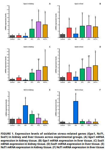
This study aimed to investigate the modulatory effects of vitamin C and vitamin E on fluorosis-induced gene expression alterations in oxidative stress (Glutathione Peroxidase 1, Neutrophil Cytosolic Factor 1, Superoxide Dismutase 1), apoptosis (Caspase 3, Caspase 8, Caspase 9), and Phosphoinositide 3–Kinase/ Protein Kinase B/ Mechanistic Target of Rapamycin signaling pathways (Phosphoinositide 3–Kinase, Protein Kinase B, Receptor Tyrosine Kinase 2, mechanistic Target of Rapamycin, Tumor Protein p53) in rats. Forty-eight male Wistar Albino rats were randomLy allocated into six groups. Sodium Fluoride (150 mg·kg–1·day–1) was administered via drinking water for 16 weeks, followed by oral vitamin supplementation (C and E) for 4 weeks. mRNA expression levels were analyzed using RT-qPCR. Sodium Fluoride exposure increased Neutrophil Cytosolic Factor 1 expression in both kidney and liver tissues (P<0.001), while reducing Glutathione Peroxidase 1 and Glutathione Peroxidase 1 levels. Vitamin E, vitamin C, and their combination significantly suppressed Neutrophil Cytosolic Factor 1 expression (P<0.001), whereas the recovery of Glutathione Peroxidase 1 and Glutathione Peroxidase 1 was significant only in kidney tissues (P<0.05). Sodium Fluoride also markedly upregulated the apoptotic genes Caspase 3, Caspase 8, and Caspase 9 (P<0.05), and antioxidant treatments substantially attenuated these increases (P<0.0001). Furthermore, all Phosphoinositide 3–Kinase/Protein Kinase B pathway-related genes were strongly overexpressed following Sodium Fluoride administration (P<0.0001), and vitamin supplementation effectively reduced these elevations in both tissues (P<0.0001). Vitamin C and vitamin E, particularly in combination, exhibit substantial protective effects by restoring gene expression patterns toward homeostatic levels. This implies that a combination of antioxidant supplements may provide a promising therapeutic tool in combating fluorosis-induced cellular dysfunction.
Downloads
References
Xiong X, Liu J, He W, Xia T, He P, Chen X, Yang K, Wang A. Dose-effect relationship between drinking water fluoride levels and damage to liver and kidney functions in children. Environ. Res. [Internet]. 2007; 103(1):112–116. doi: https://doi.org/b6fsxc DOI: https://doi.org/10.1016/j.envres.2006.05.008
Agalakova NI, Nadei OV. Inorganic fluoride and functions of brain. Crit. Rev. Toxicol. [Internet]. 2020; 50(1):28–46. doi: https://doi.org/rbc2 DOI: https://doi.org/10.1080/10408444.2020.1722061
Peng W, Xu S, Zhang J, Zhang Y. Vitamin C Attenuates Sodium Fluoride-Induced Mitochondrial Oxidative Stress and Apoptosis via Sirt1–SOD2 Pathway in F9 Cells. Biol. Trace Elem. Res. [Internet]. 2019; 191:189–198. doi: https://doi.org/rbcj DOI: https://doi.org/10.1007/s12011-018-1599-0
Strunecka A, Strunecky O. Mechanisms of Fluoride Toxicity: From Enzymes to Underlying Integrative Networks. Appl. Sci. [Internet]. 2020; 10(20):7100. doi: https://doi.org/g9hbtd DOI: https://doi.org/10.3390/app10207100
Usta A, Öner AC, Yüksek V, Dede S, Çetin S. Effect of vitamins C and E on inflammation gene expression in kidney tissue of rats with experimental fluorosis. Van Sağlık Bil. Der. [Internet]. 2021; 14(2):199–208. doi: https://doi.org/rbcm DOI: https://doi.org/10.52976/vansaglik.872528
Dede S, Öner AC, Yüksek V, Çetin S, Usta A. Investigation of the effect of vitamin C and vitamin E on serum protein fractions in experimentally induced fluorosis rats. Kocatepe Vet. J. [Internet]. 2021; 14(2):262–267. doi: https://doi.org/gj9vjd DOI: https://doi.org/10.30607/kvj.826435
Pal P, Jha NK, Pal D, Jha SK, Anand U, Gopalakrishnan AV, Dey A, Mukhopadhyay PK. Molecular basis of fluoride toxicities: Beyond benefits and implications in human disorders. Genes Dis. [Internet]. 2023; 10(4):1470–1493. doi: https://doi.org/rbcn DOI: https://doi.org/10.1016/j.gendis.2022.09.004
Kartlaşmış K, Kökbaş U, Kayrın L. Biochemistry of apoptosis. Arşiv Kaynak Tarama Derg. [Internet]. 2016; 25(1):52–69. doi: https://doi.org/rbcp DOI: https://doi.org/10.17827/aktd.16717
Fu X, Xie FN, Dong P, Li QC, Yu GY, Xiao R. High-Dose fluoride ımpairs the properties of human embryonic stem cells via JNK Signaling. Plos One [Internet]. 2016; 11(2):e0148819. doi: https://doi.org/f8rhc5 DOI: https://doi.org/10.1371/journal.pone.0148819
Fan B, Yu Y, Zhang Y. PI3K-Akt1 expression and its significance in liver tissues with chronic fluorosis. Int. J. Clin. Exp. Pathol. [Internet]. 2015 [cited 12 Dec 2025]; 8(2):1226–1236. PMID: 25973007. Available in: https://goo.su/Pin1uU
Laplante M, Sabatini DM. mTOR signaling in growth control and disease. Cell [Internet]. 2012; 149(2):274–293. doi: https://doi.org/ghxrn4 DOI: https://doi.org/10.1016/j.cell.2012.03.017
Ortiz-Barroso G, Ramírez-Orozco RE, Esparza-Villalpando V, Macedo-Mendoza M, Barrios-García T, Pulido-Hornedo NA. Antioxidants against oxidative stress induced by sodium fluoride toxicity in murine models: A systematic review. J.Trace Elem. Med. Biol. [Internet]. 2025; 88:127619. doi: https://doi.org/rbcq DOI: https://doi.org/10.1016/j.jtemb.2025.127619
Yilmaz B, Erkan M. Effects of vitamin C on sodıum fluoride- induced oxidative damage in sertoli cells. Fluoride. [Internet]. 2015 [cited 12 Dec 2025]; 48(3):241–251. Available in: https://goo.su/cYOz6
Traber MG, Stevens JF. Vitamins C and E: beneficial effects from a mechanistic perspective. Free Radic. Biol. Med. [Internet]. 2011; 51(5):1000–1013. doi: https://doi.org/bk7v9z DOI: https://doi.org/10.1016/j.freeradbiomed.2011.05.017
Li Y, Schellhorn HE. New developments and novel therapeutic perspectives for vitamin C. J. Nutr. [Internet]. 2007; 137(10):2171–2184. doi: https://doi.org/gfkcbm DOI: https://doi.org/10.1093/jn/137.10.2171
Chomczynski P, Mackey K. Modification of the TRI reagent procedure for isolation of RNA from polysaccharide – and proteoglycan-rich sources. Biotechniques [Internet]. 1995 [cited 12 Dec 2025]; 19(6):942–945. Available in: https://goo.su/Flnq
Bustin SA. A-Z of Quantitative PCR. IUL Biotechnology series. No. 5. California (USA): International University Line. 2004.
Kong H, He Z, Li H, Xing D, Lin J. The Association between fluoride and bone mineral density in US children and adolescents: A pilot study. Nutrients [Internet]. 2024; 16(17):2948. doi: https://doi.org/rbcr DOI: https://doi.org/10.3390/nu16172948
Lavanya S, Hdema-Shree K, Ramani P. Fluoride effect on renal and hepatic functions: A comprehensive decade review of In vitro and In vivo studies. J. Oral Biol. Craniofac. Res. [Internet]. 2024; 14(6):735–745. doi: https://doi.org/rbcs DOI: https://doi.org/10.1016/j.jobcr.2024.10.002
Subhashree T, Yashoda R, Manjunath PP. Neurotoxicity of fluoride: A narrative review. Int. J. Med. Sci. Dent. Res. [Internet]. 2024 [cited 11 Nov 2025]; 7(5):59–66. Available in: https://goo.su/rBETV
Basha MP, Sujitha NS. Chronic fluoride toxicity and myocardial damage: antioxidant offered protection in second generation rats. Toxicol. Int. [Internet]. 2011; 18(2):99–104. doi: https://doi.org/dwxrpt DOI: https://doi.org/10.4103/0971-6580.84260
Chinoy N, Sharma A. Amelioration of fluoride toxicity by vitamins E and D İn reproductive functions of male mice. Fluoride [Internet]. 1998 [cited 15 Nov 2025]; 31(4):203–216. Available in: https://goo.su/uxO2C
Azab A, Albasha M, Jbireal J, Adwas A. Sodium fluoride induces hepato-renal oxidative stress and pathophysiological changes in experimental animals. Open J. Apoptosis [Internet]. 2018; 7:1–23. doi: https://doi.org/rbct DOI: https://doi.org/10.4236/ojapo.2018.71001
Yüksek V, Çetin S, Usta A. The effect of vitamin E and selenium combination in repairing fluoride-induced DNA damage to NRK–52E cells. Mol. Biol. Rep. [Internet]. 2020; 47(10):7761– 7770. doi: https://doi.org/rbcv DOI: https://doi.org/10.1007/s11033-020-05852-2
Shao D, Zhang J, Tang L, Yu Q, Hu X, Ruan Q, Ouyang W, Zhang Z. Effects and molecular mechanism of L-type calcium channel on fluoride-induced kidney injury. Biol. Trace Elem. Res. [Internet]. 2020; 197(1):213–223. doi: https://doi.org/rbcw DOI: https://doi.org/10.1007/s12011-019-01987-x
Guo L, Yang B, Chen F, Yuan X, Cheng J, Chen X, Zhou Y, Yang X, Li Y, Liu Y, Tang D, Wang F. Disruption of the SIRT1/PI3K/ AKT signaling axis mediates fluoride-induced cardiotoxicity: Evidence from in vitro and Zebrafish models. Biol. Trace Elem. Res. [Internet]. 2026; 204(4):2411–2426. doi: https://doi.org/rbcx DOI: https://doi.org/10.1007/s12011-025-04828-2
Ma L, Zhang R, Li D, Qiao T, Guo X. Fluoride regulates chondrocyte proliferation and autophagy via PI3K/AKT/mTOR signaling pathway. Chem. Biol. Interact. [Internet]. 2021; 349:109659. doi: https://doi.org/rbcz DOI: https://doi.org/10.1016/j.cbi.2021.109659
Öner AC, Dede S, Yur F, Öner A. The effect of vitamin C and vitamin E on DNA damage, oxidative status, and some biochemical parameters in rats with experimental fluorosis. Fluoride [Internet]. 2020 [cited 11 Jan 2026]; 53(1–2):154–163. Available in: https://goo.su/EJHzK