Determinación de los rangos de referencia de la escala vertebral cardíaca en Gacelas subgutturosa
Resumen
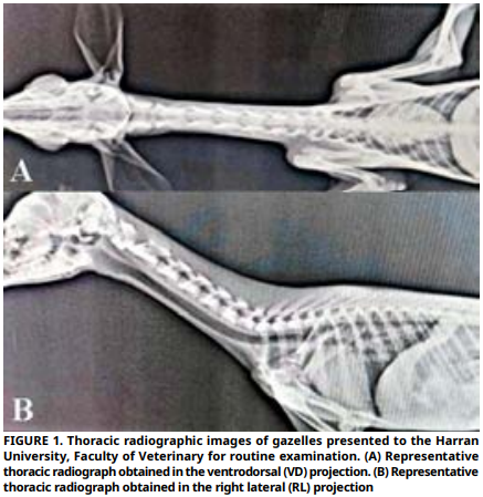
Los datos de referencia por especie, corresponden a la evaluación radiográfica de las estructuras cardíacas en gacelas (Gazella subgutturosa) son limitados; por lo tanto, este estudio prospectivo de referencia anatómica, la cual tuvo como objetivo determinar los valores de la escala cardíaca vertebral en gacelas jóvenes clínicamente sanas y evaluar los posibles efectos de la posición del cuello, el sexo y la edad en estas mediciones. Se incluyeron en el estudio veinte gacelas sanas, con edades comprendidas entre 0 y 6 meses (10 hembras y 10 machos). En todos los individuos se obtuvieron radiografías torácicas en proyección lateral derecha y ventrodorsal durante la fase de inspiración completa. Los ejes largo y corto del corazón se midieron de forma digital, y la suma de estas mediciones escala cardíaca vertebral se expresó en unidades vertebrales, utilizando la cuarta vértebra torácica como punto de referencia. Los valores medios de escala cardíaca vertebral fueron de 9,59 ± 0,59 unidades vertebrales en la proyección lateral derecha y de 9,61 ± 0,58 unidades vertebrales en la proyección ventrodorsal, sin diferencias estadísticamente significativas entre ambas posiciones (P=0,494). La medición del eje largo fue significativamente mayor en la proyección ventrodorsal en comparación con la lateral derecha (P=0,001); sin embargo, esta diferencia no se reflejó en el valor total de la escala cardíaca vertebral. No se detectaron diferencias significativas entre sexos en cuanto a la escala cardíaca vertebral ni en las mediciones de los ejes cardíacos (P>0,05). La repetibilidad de las mediciones fue elevada y la concordancia interobservador se consideró excelente (Coeficiente de Correlación Intraclase = 0,91; IC del 95 %: 0,85– 0,96; P<0,001). Los valores medios de escala cardíaca vertebral obtenidos fueron similares a los rangos de referencia descritos en perros (9,7 ± 0,5 unidades vertebrales), sin evidenciarse diferencias estadísticamente significativas. En conclusión, este estudio constituye el primer reporte que define los rangos de referencia de la escala vertebral cardíaca en gacelas subgutturosa y proporciona un criterio objetivo para la evaluación radiográfica de la silueta cardíaca, contribuyendo de manera significativa al diagnóstico clínico y al seguimiento cardiológico en especies de rumiantes exóticos.
Descargas
Citas
Buchanan JW, Bücheler J. Vertebral scale system to measure canine heart size in radiographs. J. Am. Vet. Med. Assoc. [Internet]. 1995 [cited 12 Oct 2025]; 206(2):194–199. PMID: 7751220. Available in: https://goo.su/OodSBo DOI: https://doi.org/10.2460/javma.1995.206.02.194
O’Leary CA, Mackay BM, Taplin RH, Atwell RB. Echocardiographic parameters in 14 healthy English bull terriers. Aust. Vet. J. [Internet]. 2003; 81(9):535–542. doi: https://doi.org/dg54fw DOI: https://doi.org/10.1111/j.1751-0813.2003.tb12881.x
Root CR, Bahr RJ. The heart and great vessels. In: Thrall DE, editor. Textbook of Veterinary Diagnostic Radiology. 4th ed. Philadelphia (USA): Saunders; 2002. p. 402–419.
Ghadiri A, Avizeh R, Rasekh A, Yadegari A. Radiographic measurement of vertebral heart size in healthy stray cats. J. Feline Med. Surg. [Internet]. 2008; 10(1):61–65. doi: https://doi.org/bc9x94 DOI: https://doi.org/10.1016/j.jfms.2007.06.015
Litster AL, Buchanan JW. Vertebral scale system to measure heart size in radiographs of cats. J. Am. Vet. Med. Assoc. [Internet]. 2000; 216(2):210–214. doi: https://doi.org/b9hq7w DOI: https://doi.org/10.2460/javma.2000.216.210
Stepien RL, Benson KG, Forrest LJ. Radiographic measurement of cardiac size in normal ferrets. Vet. Radiol. Ultrasound [Internet]. 1999; 40(6):606–610. doi: https://doi.org/bfvjtr DOI: https://doi.org/10.1111/j.1740-8261.1999.tb00886.x
Ukaha RO, Kene RO, Gboniko OE. Vertebral scale system to measure heart size in thoracic radiographs of West African dwarf goats. Niger. Vet. J. [Internet]. 2013 [cited 12 Oct 2025]; 34(4):912–916. Available in: https://goo.su/PYMIug
Onuma M, Ono S, Ishida T, Shibuya H, Sato T. Radiographic measurement of cardiac size in 27 rabbits. J. Vet. Med. Sci. [Internet]. 2010; 72(4):529–531. doi: https://doi.org/bvnsks DOI: https://doi.org/10.1292/jvms.09-0390
Nelson NC, Mattoon JS, Anderson DE. Radiographic appearance of the thorax of clinically normal alpaca crias. Am. J. Vet. Res. [Internet]. 2011; 72(11):1439–1448. doi: https://doi.org/btd2sj DOI: https://doi.org/10.2460/ajvr.72.11.1439
Bavegems V, Van Caelenberg A, Duchateau L, Sys SU, Van Bree H, De Rick A. Vertebral heart size ranges specific for whippets. Vet. Radiol. Ultrasound [Internet]. 2005; 46(5):400–403. doi: https://doi.org/c4r6qw DOI: https://doi.org/10.1111/j.1740-8261.2005.00073.x
Hansson K, Häggström J, Kvart C, Lord P. Interobserver variability of vertebral heart size measurements in dogs with normal and enlarged hearts. Vet. Radiol. Ultrasound [Internet]. 2005; 46(2):122–130. doi: https://doi.org/fj2vpm DOI: https://doi.org/10.1111/j.1740-8261.2005.00024.x
Lamb CR, Tyler M, Boswood A, Skelly BJ, Cain M. Assessment of the value of the vertebral heart scale in the radiographic diagnosis of cardiac disease in dogs. Vet. Rec. [Internet]. 2000; 146(24):687–690. doi: https://doi.org/fvrw37 DOI: https://doi.org/10.1136/vr.146.24.687
Demircioğlu I, Gezer-Ince N. Three-dimensional modeling of computed tomography images of limb bones of gazelles (Gazella subgutturosa). Anat. Histol. Embryol. [Internet]. 2020; 49(6):695–707. doi: https://doi.org/g6j2bk DOI: https://doi.org/10.1111/ahe.12564
Fadakar D, Bärmann EV, Lerp H, Mirzakhah M, Naseri-Nasari M, Rezaei HR. Diversification and subspecies patterning of the goitered gazelle (Gazella subgutturosa) in Iran. Ecol. Evol. [Internet]. 2020; 10(12):5877–5891. doi: https://doi.org/qs8n DOI: https://doi.org/10.1002/ece3.6324
Hussein MF, Aljumaah RS, Alshaikh MA, Elnabi AG, Sandouka MA, Homeida A. Coagulation parameters of captive mountain gazelle (Gazella gazella Pallas, 1766; Bovidae: Antilopinae) and Nubian ibex (Capra ibex nubiana Cuvier, 1825; Bovidae: Caprinae). Comp. Clin. Pathol. [Internet]. 2012; 21:527–531. doi: https://doi.org/bvwv5g DOI: https://doi.org/10.1007/s00580-010-1124-0
Tharwat M, Al-Sobayil F, Al-Hawas A, Buczinski S. Elevated serum concentration of cardiac troponin I in a Dorcas gazelle (Gazella dorcas) with mitral vegetation. Comp. Clin. Pathol. [Internet]. 2014; 23:469–473. doi: https://doi.org/qs8p DOI: https://doi.org/10.1007/s00580-013-1847-9
Taylor CJ, Simon BT, Stanley BJ, Lai GP, Thieman-Mankin KM. Norwich terriers possess a greater vertebral heart scale than the canine reference value. Vet. Radiol. Ultrasound [Internet]. 2020; 61(1):10–15. doi: https://doi.org/g8cr93 DOI: https://doi.org/10.1111/vru.12813
Bodh D, Hoque M, Saxena AC, Gugjoo MB, Bist D, Chaudhary JK. Vertebral scale system to measure heart size in thoracic radiographs of Indian Spitz, Labrador Retriever and mongrel dogs. Vet. World [Internet]. 2016; 9(4):371–376. doi: https://doi.org/qs8q DOI: https://doi.org/10.14202/vetworld.2016.371-376
Babicsak VR, Alves LS, Tsunemi MH, Vulcano LC. Radiographic measurements of heart size in young female Bergamasca sheep. Pesq. Vet. Bras. [Internet]. 2017; 37(12):1526–1530. doi: https://doi.org/g74jgr DOI: https://doi.org/10.1590/s0100-736x2017001200027
Dias S, Anselmi C, Espada Y, Martorell J. Vertebral heart score to evaluate cardiac size in thoracic radiographs of 124 healthy rats (Rattus norvegicus). Vet. Radiol. Ultrasound [Internet]. 2021; 62(4):394–401. doi: https://doi.org/qs8r DOI: https://doi.org/10.1111/vru.12973
Della -Torre PK , Kirb y A C , C hu r ch DB , Ma lik R. Echocardiographic measurements in greyhounds, whippets and Italian greyhound dogs with similar conformation but different size. Aust. Vet. J. [Internet]. 2000; 78(1):49–55. doi: https://doi.org/bnhfwb DOI: https://doi.org/10.1111/j.1751-0813.2000.tb10361.x
Marin LM, Brown J, McBrien C, Baumwart R, Samii VF, Couto CG. Vertebral heart size in retired racing greyhounds. Vet. Radiol. Ultrasound [Internet]. 2007; 48(4):332–334. doi: https://doi.org/cg6jqp DOI: https://doi.org/10.1111/j.1740-8261.2007.00252.x
Godart PO, Ruel Y, Bertal M, Esmieu S, Gouni V, Agoulon A, Gaillot H. Accuracy of the modified vertebral heart score and the cardio-vertebral ratio for radiographic evaluation of cardiomegaly in ferrets. Vet. Radiol. Ultrasound [Internet]. 2023; 64(2):173–182. doi: https://doi.org/qs8s DOI: https://doi.org/10.1111/vru.13185
Onuma M, Kondo H, Ono S, Ueki M, Shibuya H, Sato T. Radiographic measurement of cardiac size in 64 ferrets. J. Vet. Med. Sci. [Internet]. 2009; 71(3):355–358. doi: https://doi.org/ddp7jq DOI: https://doi.org/10.1292/jvms.71.355
Rungpupradit J, Sutthigran S. Comparison between conventional and applied vertebral heart score methods to evaluate heart size in healthy Thai domestic shorthair cats. Thai. J. Vet. Med. [Internet]. 2020; 50(4):459–465. doi: https://doi.org/qs8t DOI: https://doi.org/10.56808/2985-1130.3049