Irrigación Arterial del bazo en ovejas Kangal: estudio mediante microscopia electrónica de barrido
Resumen
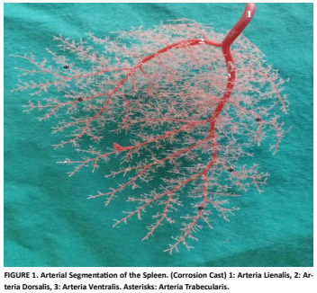
Este estudio se realizó con el objeto de demostrar la irrigación arterial del bazo, en ovejas Kangal del tipo Akkaraman, mediante microscopía electrónica de barrido y microscopía óptica. Para ellos, se utilizaron los bazos de 10 ovejas Kangal, obtenidos de una sala de faenado local. El protocolo necesario para la obtención de imágenes a partir de un microscopio electrónico, se aplicó a cinco de los bazos. Después del estudio de los tejidos, los cinco bazos restantes se tiñeron con la técnica de triple tinción de Mallory y se examinaron bajo un microscopio óptico. Macroscópicamente, se vió que el bazo tenía forma de hoja y que la arteria esplénica se dividió en dos ramas principales, la arteria dorsal y la arteria ventral. Se encontró que muchas arterias trabeculares se separaron de las ramas principales y que estas se dividieron en las arterias centrales. Estos vasos continuaron con la arteria peniciladas, cuyos extremos terminaron ciegamente en forma de bulbos. Los hallazgos obtenidos del examen con microscopio óptico fueron paralelos a los obtenidos del análisis con microscopio electrónico de barrido. Se discutieron los hallazgos obtenidos con ambos tipos de microscopia, teniendo en cuenta la literatura disponible. Los hallazgos del estudio ofrecen una guía para la intervención quirúrgica del bazo. Además, proporciona a los investigadores la información necesaria para comparar estudios que se han realizado en otras especies. Este estudio aborda la deficiencia en la literatura sobre el tema.
Descargas
Citas
Colville TP, Bassert JM. Clinical anatomy and physiology for veterinary technicians. 3th ed. Canada: Elsevier Health Sciences; 2015.
König HE, Liebich HG. Veterinary anatomy of domestic animals: Textbook and colour atlas. 7th ed. New York: Georg Thieme Verlag Stuttgart; 2020.
Aspinall V, Cappello M. Introduction to animal and veterinary anatomy and physiology. 4th ed. Wallingford, Oxfordshire, London,UK: CAB International Nosworthy Way; 2020 DOI: https://doi.org/10.1079/9781789241150.0000
Chadburn A. The spleen: anatomy and anatomical function. Semin. Hematol. [Intermet]. 2000; 37(Suppl. 1):13-21. doi: https://doi.org/bkd5dn DOI: https://doi.org/10.1016/S0037-1963(00)90113-6
Özüdoğru Z, Özdemir D, Balkaya H, Kara H. Konya Merinosunun Arteria Celiaca›sı ve Dalları Üzerine Makroanatomik Bir Çalışma. Atatürk Univ. Vet. Bilim. Derg. [Internet]. 2019; 14(1):45-51. doi: https://doi.org/pdgp DOI: https://doi.org/10.17094/ataunivbd.425567
Dursun N. Veterinary Anatomy II. 10th ed. Medisan Yayinevi, Ankara: Medisan press.; 1996.[7] Atalar O, Yılmaz
S. The branches of the arteria celiaca in the porcupine (Hystrix cristata). Vet. Med. [Internet]. 2004;49(2):52-56. doi: https://doi.org/pdg3 DOI: https://doi.org/10.17221/5675-VETMED
Yılmaz S, Atalar O, Aydın A. The branches of the arteria celiaca in badger. Indian Vet. J. [Internet]. 2004; 81:183- 187.
Getty R, Grossman JD, Rosenbaum CE, Ghoshal N, HillmanD. Sisson and Grossman’s the Anatomy of the Domestic Animals. 5th ed. Philadelphia: WB Saunders company; 1975.
Özcan S, Kürtül I, Aslan K. Arterial vascularization of the stomach in the German shepherd dog. İstanbul Univ. Vet. Fak. Derg. [Internet]. 2001[Cited 1 Dec 2024]; 27(2):487-494. Available in: https://goo.su/SfHqnvF
Gupta SC, Gupta CD, Gupta SB. Arterial segmentation in the goat (Capra hircus) spleen: A study by corrosion cast. Acta Anat. [Internet] 1978; 102(1):102-104. doi: https://doi.org/dbzw74 DOI: https://doi.org/10.1159/000145625
Gupta SC, Gupta CD, Gupta SB. Arterial segmentation in the spleen of the sheep (Ovis aries). J. Anat. [Internet]. 1979 [cited Nov 12 2024]; 129(Pt 2):257-260. Available in: https://goo.su/0o0Gcdq
Rossi Feliciano MA, Da Câmara Barros FFP, Coutinho LN, De Brito MBS, Uscategui RR, Santos VJC, De Almeida VT, Kawanami AE, Nociti RP, Fernandez Machado MR, Russiano Vicente WR. Conventional and Doppler abdominal ultrasonography in pacas (Cuniculus paca). Acta Sci. Vet. [Internet]. 2014 [Accessed Dec 1 2024]; 42(1):1-6. Available in: https://goo.su/vGbm
Nawal AN, Maher MA. Gross anatomical, radiographic and ultra-structural identification of splenic vasculature in some ruminants (camel, buffalo calf, sheep and goat). Int. J. Adv. Res. Biol. Sci. [Internet]. 2018 [cited Dec 02 2024]; 5(2):44-65. Available in: https://goo.su/F8eui
Thacker C, Korn R, Millstine J, Harvin H, Van Lier Ribbink JA, Gotway MB. Sclerosing angiomatoid nodular transformation of the spleen: CT, MR, PET, and 99mTc-sulfur colloid SPECT CT findings with gross and histopathological correlation. Abdom. Imaging. [Internet]. 2010; 35(6):683-689. doi: https://doi.org/fvpg2g DOI: https://doi.org/10.1007/s00261-009-9584-x
Uršıč M, Ravnik D, Hribernik M, Pečar J, Butinar J, Fazarinc G. Gross anatomy of the portal vein and hepatic artery ramifications in dogs: corrosion cast study. Anat. Histol. Embryol. [Internet]. 2007; 36(2):83-87. doi: https://doi.org/bpxhcr DOI: https://doi.org/10.1111/j.1439-0264.2006.00719.x
Peres Ferraz De Melo A, De Souza WM, Rodrigues RF, Alves FR, Rici REG, Guerra RR, Favaron P O, Miglino MA, Di Dio LJA. Anatomical description of arterial segments of the spleen of deer. Anat. Histol. Embryol. [Internet]. 2011; 40(4):243-248. doi: https://doi.org/fbpsbt DOI: https://doi.org/10.1111/j.1439-0264.2011.01063.x
Kırbas G, Dalga S, Akbulut Y, Aslan K. Kızıl Tilkilerde (Vulpes vulpes) Arteria Celiaca ve Dalları Üzerinde Bir Çalışma. Harran Univ. Vet. Fak. Derg. [Internet]. 2019; 8(2):168-172. doi: https://doi.org/pdnx DOI: https://doi.org/10.31196/huvfd.667680
Suri S, Sasan JS, Sarma K, Chakraborty D. Comparative gross and histomorphological studies on the spleen of sheep and goat of Jammu region of India. Explor. Anim. Med. Res. [Internet]. 2017 [Accessed 1 Dec 2024]; 7(2):179-183. Available in: https://goo.su/iPceB
Gnanadevi R, Senthilkumar S, Ta K, Ramesh G. Comparative Histoarchitectural study of splenic components in sheep and goat. Int. J. Curr. Microbiol. App. Sci. [Internet]. 2019; 8(5):1387-1394. doi: https://doi.org/pdnz DOI: https://doi.org/10.20546/ijcmas.2019.805.158
Rashad E, Hussein S, Bashir DW, Sabry Z, Maher M, El- Habback H. Anatomical, histological, histochemical, scanning and transmission electron microscopic studies on water Buffalo (Bubalus Bubalis) Spleen. J. Crit. Reviews. [Internet]. 2020 [Accessed 1 Dec 2024]; 7(15):6154-6173. Available in: https://goo.su/UIdoS
Tanaka Y. Intermediate zone” of mammalian spleens: Light and electron microscopic study of three primitive mammalian species (platypus, shrew, and mole) with special reference to intrasplenic arteriovenous communication. Am. J. Anat. [Internet]. 1990; 187(4):313-337. doi: https://doi.org/ddwsc2 DOI: https://doi.org/10.1002/aja.1001870402
Alexandre-Pires G, Pais D, Esperança Pina JA. Intermediary spleen microvasculature in Canis familiaris– Morphological evidences of a closed and open type. Anat. Histol. Embryol. [Internet]. 2003; 32(5):263-270. doi: https://doi.org/b9c775 DOI: https://doi.org/10.1046/j.1439-0264.2003.00469.x
Motta P, Murakamı T, Fujita H. Scanning electron microscopy of vascular casts: methods and applications. 1st ed. New York: Springer Science & Business Media, LLC; 1992. DOI: https://doi.org/10.1007/978-1-4615-3488-4
Bennoune O, Al-Samarrae N. Splenic arterial tree and its application in biopsy and splenectomy in camels (Camelus dromedarius). Revue Méd. Vét. [Internet]. 2012 [cited: 1 Dec 2024]; 163(10):461-464. Available in: https://goo.su/7kO9lor
Osman F, El-Ayat M, El-Khaligi G. Parenchymal distribution of the splenic vessels in buffalo calves. Vet. Med. J. [Internet]. 1987 [cited: 1 Dec 2024]; 35(2):175-181. Available in: https://goo.su/j816m8
Bancroft JD, Gamble M. Theory and practice of histological techniques. 5th ed. Nothinghum UK: Elsevier health sciences. 2008.[28] Abu-Zaid SMS, El-Khaligi GEM, El-Nahla SMM. Some gross anatomical studies on the topography, arterial supply and venous drainage of the spleen of the one Humped camel (Camelus dromedarius). Alex. J. Vet. Sci. [Internet]. 1985 [cited 1 Dec 2024]; 1(2):45-59. Available in: https://goo.su/XI2xTe
Redmond HP, Redmond JM, Rooney BP, Duignan JP, Bouchier-Hayes DJ. Surgical anatomy of the human spleen. BJS. [Internet]. 1989; 76(2):198-201. doi: https://doi.org/fj6572 DOI: https://doi.org/10.1002/bjs.1800760230
Ocal M, Takcı L. Arterial segmentation in the spleen of the sheep. Anat. Histol. Embryol. [Internet]. 1991; 20(2):152-153. doi: https://doi.org/bdtx5r DOI: https://doi.org/10.1111/j.1439-0264.1991.tb00754.x
Liu DL, Xia S, Xu W, Ye Q, Gao Y, Qian J. Anatomy of vasculature of 850 spleen specimens and its application in partial splenectomy. Surgery [Internet]. 1996; 119(1): 27-33. doi: https://doi.org/d6hrfq DOI: https://doi.org/10.1016/S0039-6060(96)80209-1
Tosun M, Atalgin ŞH. Arterial vascularization of the spleen in Merino sheep. Turk. J. Vet. Anim. Sci. [Internet]. 2023; 47(3):194-201. doi: https://doi.org/pdpk DOI: https://doi.org/10.55730/1300-0128.4286
Cougard P, Trouilloud P, Morizot B, Gelle M, Autissier J. Study of the vascular segmentation of the spleen. Bulletin of the Association of Anatomists. [Internet].1984 [accessed 1 Dec 2024]; 68(200):27-33. Available in: https://goo.su/aznWw
Macneal WJÜ, Otani S, Patterson MB. The finer vascular channels of the spleen. Am. J. Pathol. [Internet]. 1927 [Accessed 1 Dec 2024]; 3(2):111-122.15. Available in: https://goo.su/ry3hPTg DOI: https://doi.org/10.1007/BF03400570
Schmidt E, Macdonald I, Groom A. Circulatory pathways in the sinusal spleen of the dog, studied by scanning electron microscopy of microcorrosion casts. J. Morphol. [Internet]. 1983; 178(2):111-123. doi: https://doi.org/d7jnpk DOI: https://doi.org/10.1002/jmor.1051780204
Pannarale L, Onori P, Ripani M, Gaudio E. Precapillary patterns and perivascular cells in the retinal microvasculature. A scanning electron microscope study.J. Anat. [Internet]. 1996; 188(Pt 3):693-703. Available in: https://goo.su/QiKVfa
Billur D.Investigation of Macrophage Subgroups in Rat Spl een by Using Different Morphological Techniques. J. Ankara Univ. Fac. Med. [Internet]. 2017;70(3),135-142. doi: https://doi.org/pdpx
Thanvi PK, Joshi S, Singh D. Histomorphological studies on spleen of sheep (Ovis aries). Vet. Pract. [Internet]. 2020 [accessed 1 Dec 2024]; 21(1):48-53. Available in: https://goo.su/zQT4RV